|
Example:
1. Load an image.
Choose Start, Load, URL ...
A dialog titled "Load Image" will appear.
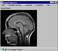
Input this URL to load a medical image (brain.gif)
https://members.tripod.com/~shamshasan786/start/brain.gif or the URL
https://members.tripod.com/~shamshasan786/start/lena256.gif for the standard Lena image.
You can also directly input a URL for loading and image from your favourite site.
Caution: The image needs to be grayscale image and its size needs to be 2n x 2n. A color image can be loaded but will not work properly, so use only grayscale image.
Click Ok in the URL dialog.
The program starts loading the image. This might take some time because of the network delay.
An information dialog will show up when the system finishes loading.
Click Ok in the information dialog.
2.Set Wavelet transform level parameters.
Choose Start, Set Level.
A dialog titled " Wavelet Transform Level" will appear.
You can set the wavelet decomposition level.
Click Ok to close the dialog.
3.Apply Shore Forward Wavelet transform.
Choose Wavelet Transform, Using Shore Wavelet, Shore Forward.
The wavelet decomposed image will be shown. 4.Apply uniform quantization.
Choose Quantizer, Uniform, Uniform Quant.
All the higher band signals fade away as they are thrown away.
5.Check the compression rate.
Choose Runlength, Run-Length.
The runlength of the image as well as compression rate will be reported.
Note: The runlength is not implimented since runlength and encoding will not work on the browser due to security restrictions.
6.Apply uniform dequantization.
Choose Quantizer, Uniform, Uniform DeQuant.
Some of the preserved high band signals will appear.
7.Apply Shore Inverse Wavelet Transform.
Choose Wavelet Transform, Using Shore Wavelet, Shore Inverse.
The decoded image will be shown. 8.Compare both images, original and decompressed.
Choose Quantizer, Comparision*1 (very little difference is observed) or Comparision*16.
|
|